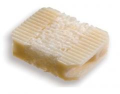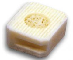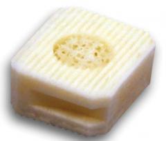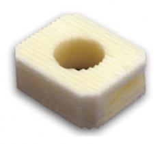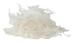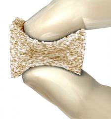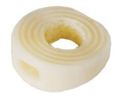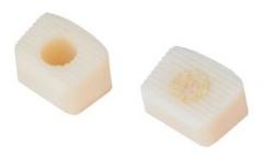
Vitoss BA Bioactive Bone Graft Substitute
Category: Biomaterials
A synthetic bone graft with bioactive glass that has a unique porosity, structure, and chemistry to help drive 3D regeneration of bone
Description
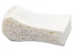
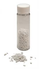
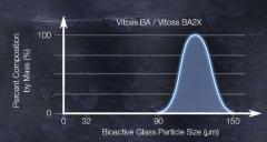
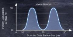
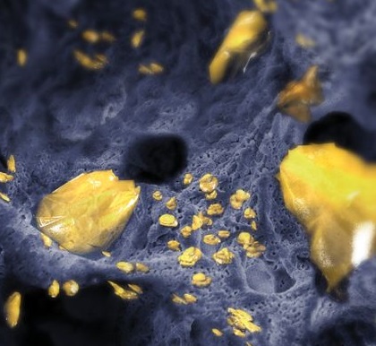
Literature has shown that bioactive glass exhibits good bonding-to-bone properties in animal models.1-3 Upon implantation, the ionic constituents (Silicon, Sodium, and Calcium) of bioactive glass are released into the surrounding environment and react with bodily fluids.4-7 This reaction produces the deposition of a thin layer of physiologic calcium phosphate at its surface, favorable for osteoblast attachment.8 This is commonly referred to as a bioactive effect, and may lead to the bonding of new bone to the scaffold.1-3,6,9-11 Vitoss BA is a highly porous calcium-phosphate (up to 90% porous)12 containing bioactive glass that is stable at physiological pH13 and resorbs during the natural remodeling process of bone.14
- The addition of bioactive glass helps create a surface favorable for osteoblast attachment1-3,6,9-11
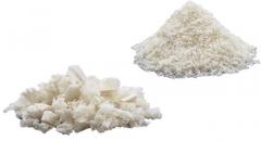
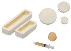
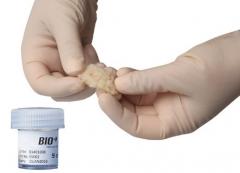
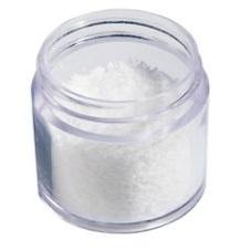
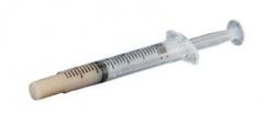
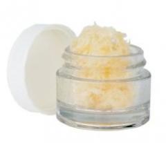
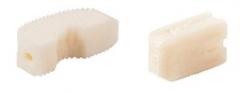
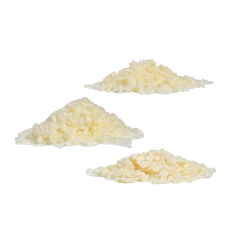
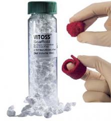
- Vitoss BA Foam Pack is stable in a fluid environment, can soak and hold bone marrow, and is moldable
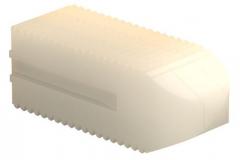
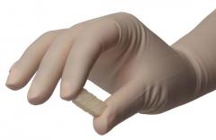
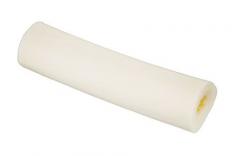
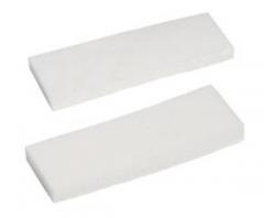
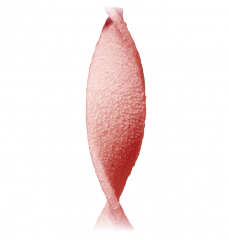
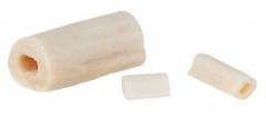
- Vitoss BA Foam Strip is flexible when wet, can soak and hold bone marrow, and is compression resistant and easy to cut
- Hench, L.L., Splinter, R.J., and Allen, W.C., Bonding Mechanisms at the Interface of Ceramic Prosthetic Materials. Journal of Biomedical Materials Research, 1971; 2(1): 117-141.
- Hench, L.L., Paschall, H.A., Direct Chemical Bond of Bioactive Glass-Ceramic Materials to Bone and Muscle. Journal of Biomedical Materials Research, 1973; 4: 25-42.
- Gross, U., The Interface of Various Glasses and Glass Ceramics with a Bony Implantation Bed. Journal of Biomedical Materials Research, 1985; 19: 251-271.
- Hench, L.L. The Story of Bioglass. Journal of Materials Science: Materials in Medicine, 2006 Nov; 17(11): 967-78.
- Oonishi, H., et al., Particulate Bioglass Compared with Hydroxyapatite as a Bone Graft Substitute. Clinical Orthopaedics and Related Research, 1997 Jan; 334: 316-25.
- Vrouwenvelder, W.C.A., Histological and Biochemical Evaluation of Osteoblasts Cultured on Bioactive Glass, Hydroxylapatite, Titanium Alloy, and Stainless Steel. Journal of Biomedical Materials Research, 1993 Apr; 27(4): 465-75.
- Xynos, I.D., Edgar, A.J., Buttery, L.D.K., Hench, L.L., and Polak, J.M., Ionic Products of Bioactive Glass Dissolution Increase Proliferation of Human Osteoblasts and Induce Insulin-like Growth Factor II mRNA Expression and Protein Synthesis. Biochemical and Biophysical Research Communications. 2000 Sep 24;276(2):461-5.
- Hench, L.L., Polak, J.M., Xynos, I.D., Buttery, L.D.K., Bioactive Materials to Control Cell Cycle. Materials Research Innovations, 2000; 3(6): 313-323.
- Sanders, D.M., Hench, L.L., Mechanisms of Glass Corrosion. Journal of American Ceramic Society.1973; 56(7): 373-377.
- Hench, L.L., Characterization of Glass Corrosion and Durability. Journal of Non-Crystalline Solids, 1975; 19: 27-39.
- Ogino, M., Hench, L.L., Formation of Calcium Phosphate Films on Silicate Glasses. Journal of Non-Crystalline Solids, 1980; 38 and 39: 673-678.
- Stryker Spine Test Report P/N 1070-0008R
- Rey C, Combes C, Drouet C, Grossin D. (2011). Bioactive Ceramics - Physical Chemistry. In Ducheyne, Paul. Comprehensive Biomaterials. 1. Elsevier. pp. 187–281.
- Anker et al, Ultraporous Beta-Tricalcium Phosphate is Well Incorporated in Small Cavitary Defects. Clinical Orthopaedics and Related Research, 2005 May; 434: 251-7.