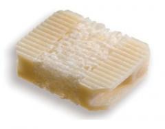
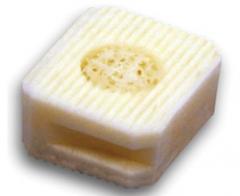
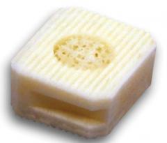
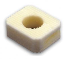
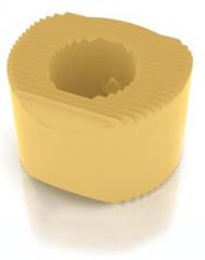
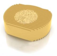
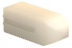
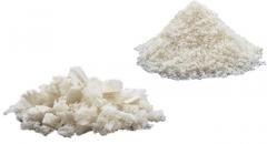
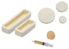
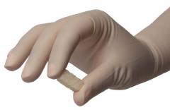
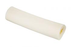
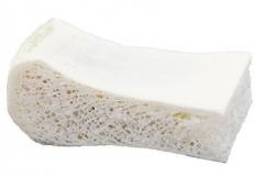
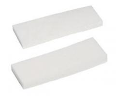
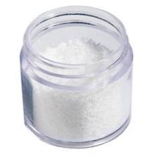
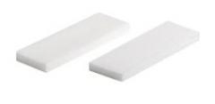
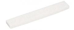
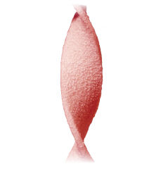
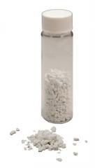
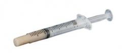
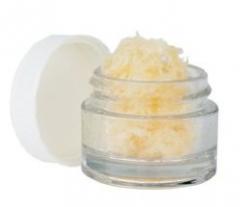
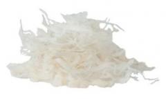
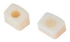
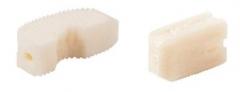
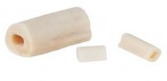
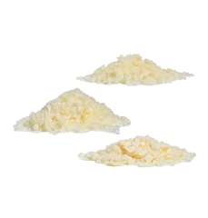
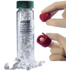
Vitoss BA Bioactive Bone Graft Substitute
Category:
Biomaterials
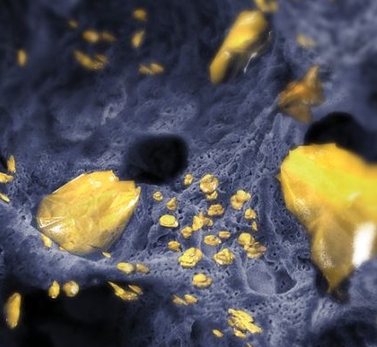
A synthetic bone graft with bioactive glass that has a unique porosity, structure, and chemistry to help drive 3D regeneration of bone
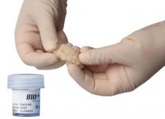
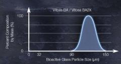
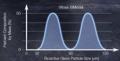
Literature has shown that bioactive glass exhibits good bonding-to-bone properties in animal models.1-3 Upon implantation, the ionic constituents (Silicon, Sodium, and Calcium) of bioactive glass are released into the surrounding environment and react with bodily fluids.4-7 This reaction produces the deposition of a thin layer of physiologic calcium phosphate at its surface, favorable for osteoblast attachment.8 This is commonly referred to as a bioactive effect, and may lead to the bonding of new bone to the scaffold.1-3,6,9-11 Vitoss BA is a highly porous calcium-phosphate (up to 90% porous)12 containing bioactive glass that is stable at physiological pH13 and resorbs during the natural remodeling process of bone.14
 Loading...